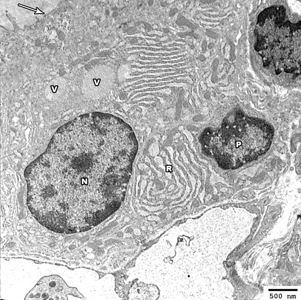
Figure 8.

Electron photomicrograph of the thyroid gland of adult rat treated with low dose of MSG (group II) showing columnar follicular cell with oval euchromatic nucleus (N) and other cell showing irregular nuclei (P) with increased condensation of its peripheral chromatin and widening of its nuclear pores. The cytoplasm of the follicular cells revealed mild dilated rER cisternea (R) and colloidal vesicles (V) are also seen above the nucleus. The apical border revealed partial loss of the projecting microvilli (arrow) (Uranyl acetate and lead citrate × 2500).
