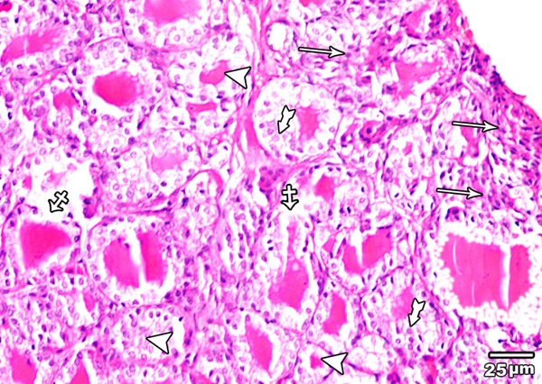
Figure 13.

A section in the thyroid gland of adult rat treated with high toxic dose of MSG (group IV) showing an area with complete loss of the normal architecture of the thyroid gland (arrow), and other with disruption of the basal laminae of some follicles with their coalescence (crossed arrow). Follicular hyperplasia (tailed arrow) and reduction in amount of colloid (arrow head) are also obvious (H&E × 400).
