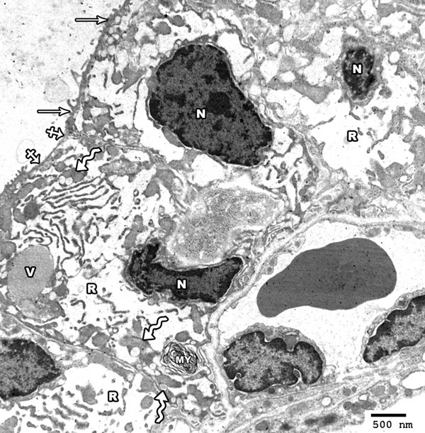
Figure 16.

Electron photomicrograph of the thyroid gland of adult rat treated with high toxic dose of MSG (group IV) showing columnar follicular cells having oval small sized irregular hyperchromatic nuclei (N). Marked dilatation of rER (R) and vacuolation of mitochondria (zigzag arrows) are seen. The cytoplasm contains colloid vacuoles (V) and a myeloid body (MY). The apical surface revealed short microvilli (arrow) with area of partial loss (crossed arrow) (Uranyl acetate and lead citrate × 2000).
