
Figure 1.
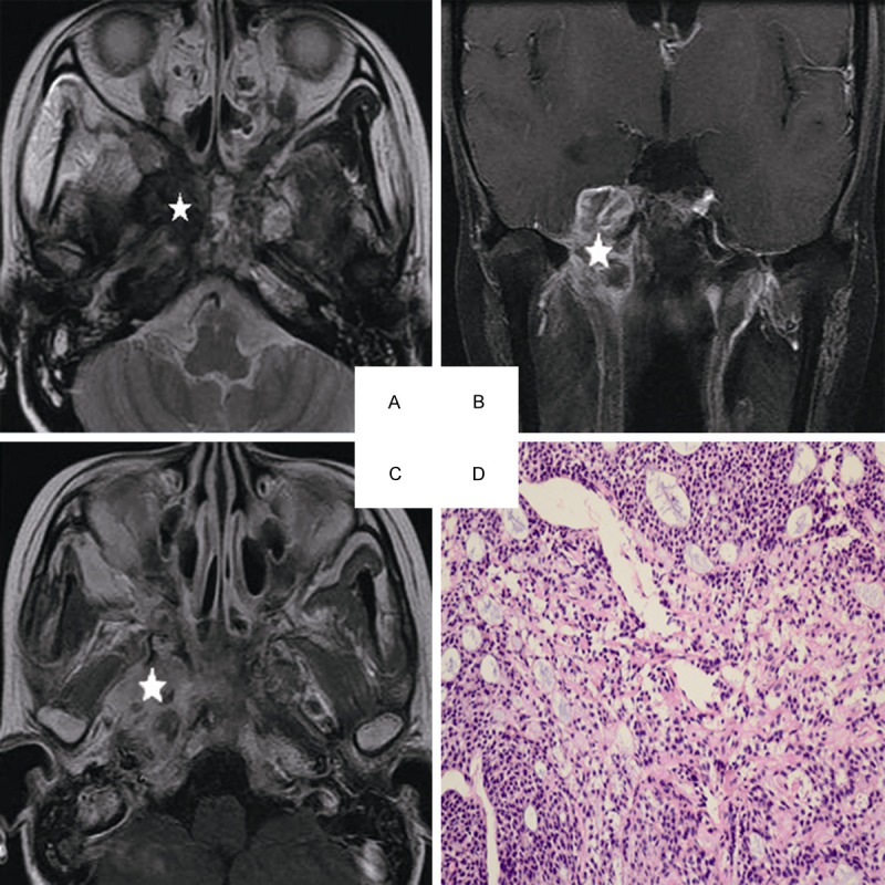
Head and neck MRI from a 25-year-old female with ACC of the nasopharynx. A. Axial plane, T2-weighted image. B. Coronal plane, T1-weighted image after contrast medium administration. C. Axial plane, T1-weighted image after contrast medium administration. D. The characteristic eosinophilic basement membrane material are displaying in pseudocysts. True gland lined by cuboidal epithelium are visible, (hematoxylin-eosin, original magnification × 200): The lesion invades to the pterygopalatine fossa, the parapharyngeal space, the pterygoid process and the skull base representing isosignal intensity on the T2-weighted image(white star), high signal intensity on the contrast-enhanced T1-weighted image (white star). There are mixed hypointense, isointense, and hyperintense in the lesion (white star).