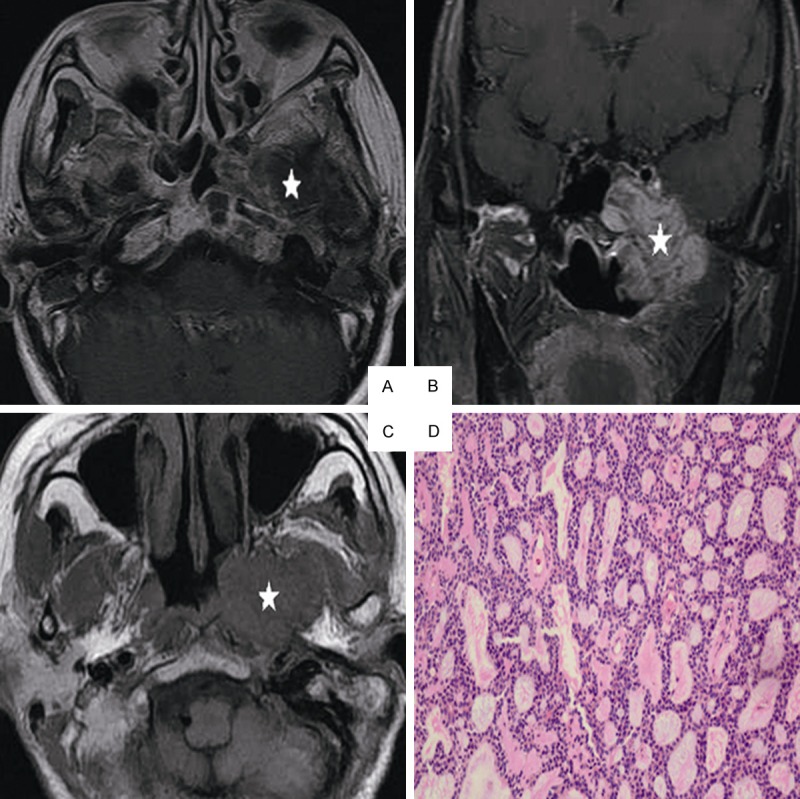
Figure 2.

Head and neck MRI from a 51-year-old female with ACC of the nasopharynx. A. Axial plane, T1-weighted image after contrast medium administration. B. Coronal plane, T1-weighted image after contrast medium administration, C. Axial plane, T1-weighted image, D. Cribriform growth pattern displaying several prominent pseudocysts surrounded by basaloid cells (hematoxylin-eosin, original magnification × 200): The lesion invades the parapharyngeal space, the sphenoid sinus, the petrous apex and the muscles on the contrast-enhanced T1-weighted image without enhancement on the image (white star). On the T1-weighted image, the lesion represents low signal intensity invading the nasal cavity, the parapharyngeal space (white star).
