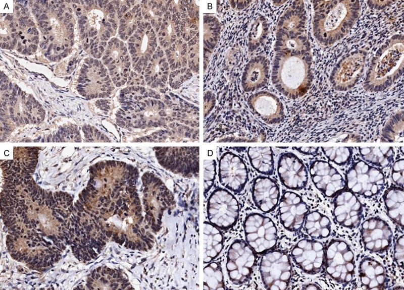
Figure 1.

Immunohistochemical staining of IFITM1 in human colorectal cancers. Representative images from immunohistochemical staining of IFITM1 in colorectal cancer and matched adjacent noncancerous tissues. Weak IFITM1 staining (A), moderate IFITM1 staining (B), and strong IFITM1 staining (C). Negative staining of IFITM1 in matched noncancerous tissues adjacent to tumors (D). Magnification: ×200.
