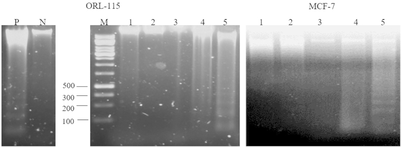
Figure 4. DNA gel electrophoresis of inter-nucleosome DNA fragmentation in 1.5% (w/v) agarose gel at 6, 12 and 24 hrs treatment in MCF-7 and ORL-115 cell lines.
Lane P: positive control with 1’-(S)-1’-acetoxychavicol acetate (ACA) treated MCF-7 cells19; Lane N: negative control (untreated MCF-7 cells); Lane M: DNA molecular weight marker; Lane 1: Untreated cells; Lane 2: PBS treated cells for 24 hrs; Lane 3: HKB treated at 6 hrs; Lane 4: HKB treated at 12 hrs; Lane 5: HKB treated at 24 hrs. DNA laddering was demonstrated in cells treated with MIP HKB in Lane 5.

