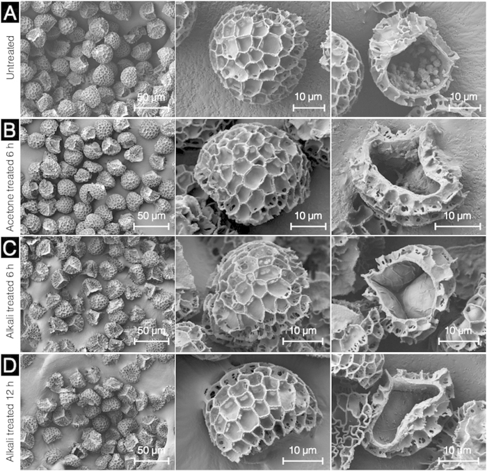
Figure 2. Characterization of L. clavatum spores during different stages of pre-acid treatment by scanning electron microscopy.
(A) Untreated L. clavatum spores are intact with uniform size, well defined microstructure, and cross section indicating sporoplasmic cellular organelles and biomolecules. (B) Defatted spores at different magnifications are intact and the cross section reveals sporoplasmic biomolecules. (C) and (D) respectively show SECs after 6 h and 12 h alkaline lysis indicating intact capsules with the cross section revealing residual proteinaceous debris and the cellulosic intine layer.

