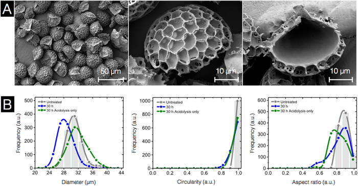
Figure 8. Characterization of 30 h acidolysis-only sporopollenin exine capsules (SECs) using scanning electron microscopy (SEM) and dynamic imaging particle analysis (DIPA).
(A) SEM images indicating an intact, well defined microstructure with a clean empty inner cavity. (B) Micromeritic properties of untreated spores and SECs. Plots are representative graphs of diameter, circularity, and aspect ratio, obtained by the spline curve fitting of histogram data from 1000 well-focused particle images from triplicate independent batches (n = 3).

