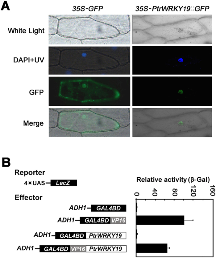
Figure 3. Subcellular localization and transactivation assays of PtrWRKY19.

Onion epidermis was transformed with 35S-PtrWRKY19::GFP and 35S-GFP constructs by particle bombardment. The position of nucleus was ensured by DAPI staining and bright-field images were compared. In this experiment, 35S-GFP was used as control. (B) Transcriptional activation analysis of PtrWRKY19 analyzed by the chimeric reporter/effector assay in yeast on the plates with solid SD medium. Data represent mean of three biological repeats ± SD. GAL4DB null vector was used as a negative control and GAL4DB fused with VP16 was used a positive control.
