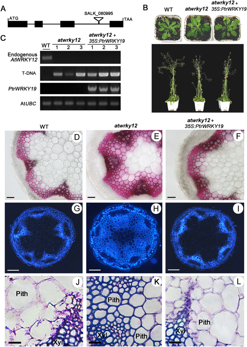
Figure 4. Complementation of the Arabidopsis wrky12 mutant with PtrWRKY19.
(A) AtWRKY12 gene structure and T-DNA insertion site. (B) Visible phenotypes of WT, wrky12 mutant and wrky12 mutant transformed with 35S-PtrWRKY19 gene. (C) Expression levels of AtWRKY12 and PtrWRKY19 in WT, wrky12 mutant and transgenic plants by PCR analysis. AtUBC gene was used as the control. (D–F) Phloroglucinol staining of the stems from WT, wrky12 mutant and wrky12 mutant complemented with 35S-PtrWRKY19 gene, respectively. (G–I) UV autofluorescence of cross-sections of the stems from WT, wrky12 mutant and wrky12 mutant complemented with 35S-PtrWRKY19 gene, respectively. (J–L) Light microscopy of pith cell walls by toluidine bule staining. Xyl, Xylem. Scale bar represent 50 μm (D–F); 100 μm (G–I); 20 μm (J–L).

