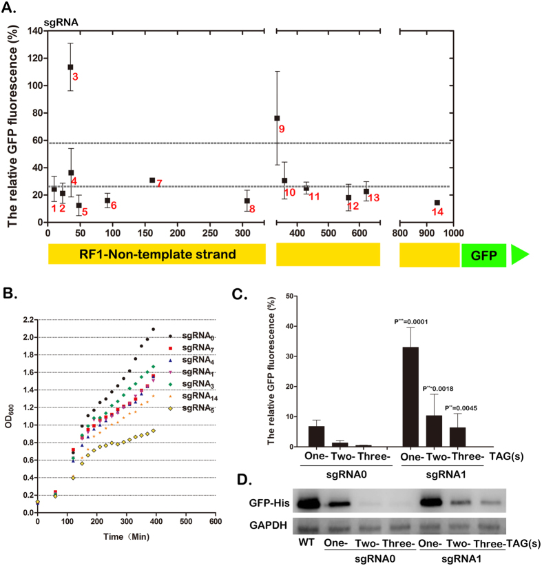
Figure 2. CRISPRi-mediated RF1 repression and the resulting effects on host cell growth and UAA incorporation into GFP protein.
(A) Comparison of the knockdown effect of 14 sgRNAs designed complementary to the different loci of RF1 gene, which was fused with GFP as a reporter. The expression level of RF1 was quantitated by GFP fluorescence (485 and 520 nm) and normalized to the control sgRNA. Error bars show one standard deviation from the mean of at least three values. (B) The growth curves of BL21 (DE3) host strains transformed with pCCR-sgRNAx. The transformed strains were separately inoculated into Luria-Bertani (LB) medium containing 35 ug/ml spectinomycin and then grew overnight at 220 rpm under 37 oC. Then the cultures were diluted to 0.1 with IPTG added to a final concentration of 0.5 mM. The OD600 values were measured every 20 minutes. (C) RF1 repression-induced incorporation enhancement of UAA into GFP gene containing one-, two- and three-TAG codons in BL21(DE3) strains. The GFP fluorescence per OD600 was measured relative to the wild-type GFP (WT) and was measured in 3 independent batches of cells. (D) Western blot analysis of the expression levels of GFP reporter, which contain one, two or three UAAs, upon knockdown of RF1 by sgRNA0 and sgRNA1, respectively, with GAPDH acted as an internal control.

