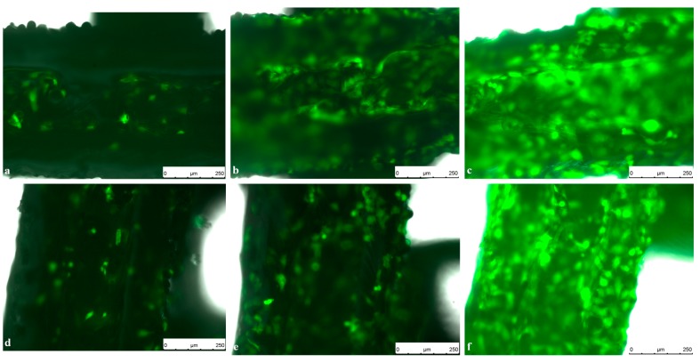
Figure 7.
Live Cell Imaging (LCI) of GFP-Osteoblasts seeded on titanium implants on (a) Day 1; (b) Day 4 and (c) Day 7 compared to GFP-Osteoblasts seeded on PCL coated titanium implants on (d) Day 1; (e) Day 4 and (f) Day 7. Pictures were taken in 10-fold magnification using an exposure time of 6 ms, a gain of 5.8 and an intensity of 3 s. Scale bar: 250 µm.

