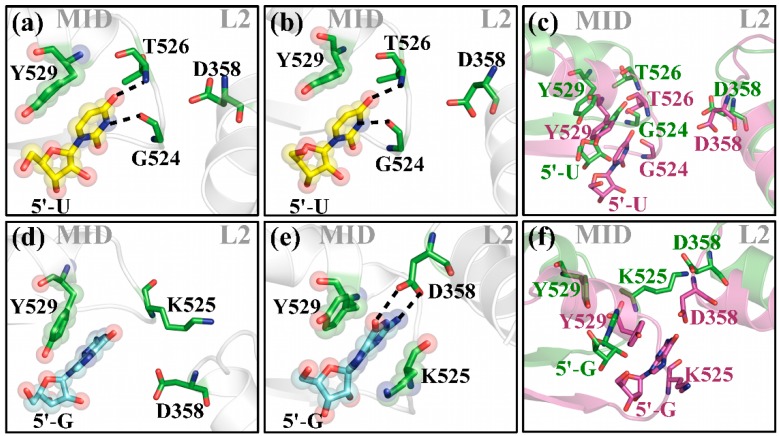
Figure 6.
Close-up of the guide RNA 5'-end and relevant protein residues of the MID domain and L2 linker region. The MID domain and the L2 helix are represented in cartoon (grey); important protein residues (green); and 5′-U (yellow); or 5′-G (blue) are shown in sticks. For clarity, merely the terminal base is shown. Hydrogen bonds are represented by black dotted lines. Transparent spheres highlight stacking interactions between protein residues and the corresponding bases. (a,d) show the initial situation; and (b,e) after 20 ns of simulation of 5′-U and 5′-G terminated hAgo2 bound guide strands, respectively; (c,f) superposition of (a,b); and (d,e) with neighboring protein residues at t = 0 ns are shown in green and at t = 20 ns in magenta.

