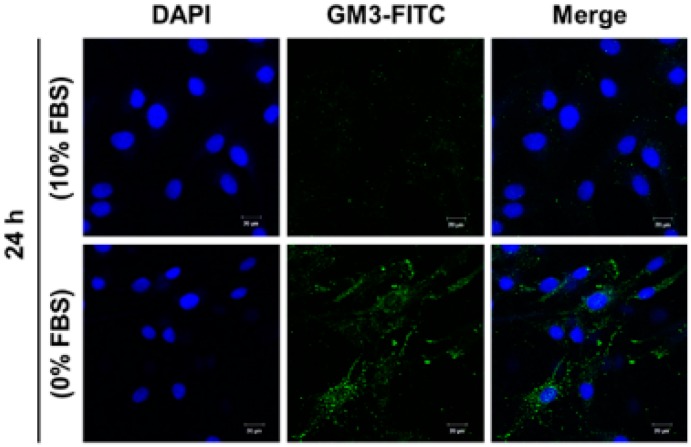
Figure 5.
Confocal analysis of ganglioside GM3 expression in SD-induced MG-63 cells. After incubation for 24 h in standard medium containing 10% FBS or 0% FBS, cells were immunostained with anti-GM3 antibodies (FITC; green). Nuclei were stained with DAPI (blue) and analyzed by confocal microscopy. Scale bar: 20 µm.

