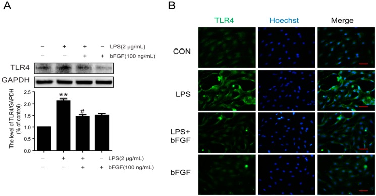
Figure 6.
Effect of bFGF on expression of TLR4 in LPS stimulated astrocytes. Cells were incubated in the presence of LPS (2 µg/mL) with or without bFGF (100 ng/mL) for 24 h. (A) Western blot of TLR4 and densitometric analyses; (B) Immunofluorescence of TLR4 (green), the nuclear is labeled by Hoechst (blue). ** p < 0.01 versus CON, # p < 0.05 versus LPS. All results represent at least three independent experiments; Scale bar is 50 µm.

