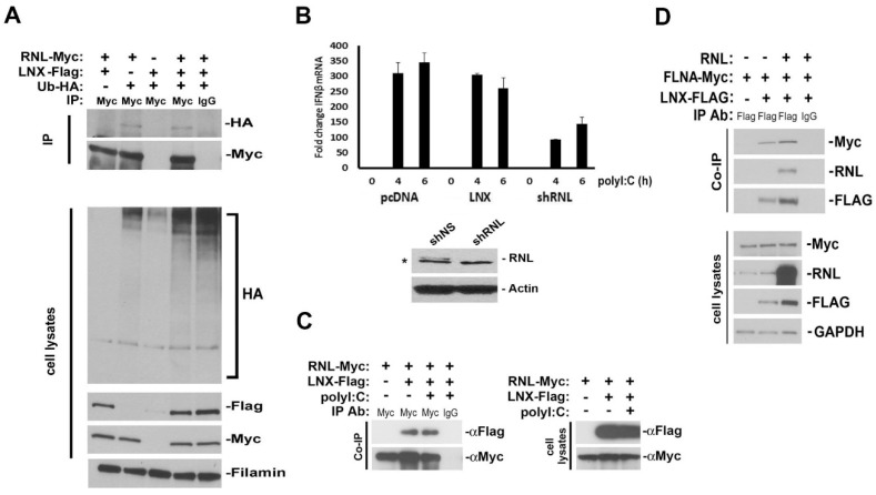
Figure 3.
Characterization and consequences of LNX expression. (A) Ubiquitylation of RNase-L is not enhanced by LNX expression. 293T cells were transfected with LNX, RNase-L, and HA-tagged ubiquitin (Ub). The presence of Ub-HA-conjugated RNase-L was determined by Western blot; (B) Hela cells were transfected with the indicated vectors. After 48 h, cells were transfected with polyI:C for 4 and 6 h and then analyzed by RT-qPCR for IFNβ expression, relative to rpl13a mRNA. Error bars represent standard deviation of two experiments. Western blot demonstrates shRNA knockdown of RNase-L expression. * indicates a non-specific band (C) 293T cells were transfected with LNX and RNase-L. After 48 h, cells were transfected with polyI:C for 6 h and then interaction was analyzed by IP; (D) Analysis of FLNA-myc and LNX-Flag interaction. FLNA and LNX were transfected into 293T cells in the presence or absence of RNase-L. Cell lysates were immunoprecipitated for LNX-Flag and Western blots probed for FLNA interaction. The presence of exogenous RNase-L did not alter the interaction.

