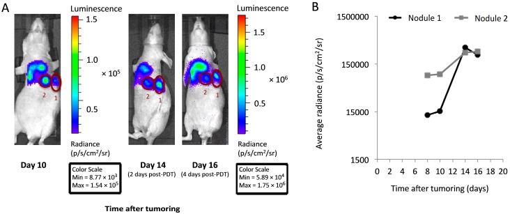
Figure 3.
In vivo imaging facilitates the identification of distinct tumor nodules with an incomplete response to PDT. (A) Bioluminescence imaging in a representative PDT-treated mouse was used to identify well-circumscribed nodules with a minimal response to PDT (labeled as “1” and “2”). To aid visualization, the day 10 image is not linked to other images for scaling purposes; note the differences in the values of the pre- and post-PDT scales (min/max) between the Day 10 and the Day 14 and 16 images; (B) Plots of bioluminescence in each of nodules “1” and “2” demonstrate persistent signal at times after PDT (treatment at 150 mW/cm, 0.5 mg/kg HPPH). For quantification in (B), all images are adjusted for equivalent scale and signal is corrected for background levels.

