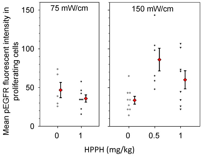
Figure 6.
PDT increases EGFR activation in the proliferating areas of intrathoracic tumors treated with a fluence rate of 150 mW/cm. Immunohistochemistry was used to quantify levels of phosphorylated EGFR (pEGFR) in areas that were positive for proliferation by Ki-67 staining. Controls received no photosensitizer (0 mg/kg HPPH), but did receive illumination at the fluence rate that corresponded to the PDT condition. Treatment of the tumor-bearing thoracic cavity was to a total dose of 50 J/cm. Open symbols (controls) and closed black symbols (treatment groups) indicate the pEGFR staining intensity in individual mice. Red diamonds indicate the mean and standard error (error bars) for each group.

