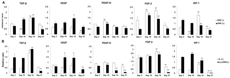
Figure 4.
(A,B) Time sequential mRNA expression of each growth factor from meniscal cells with and without either PRP or IL-1α. The changes in the mRNA levels of TGF-β, VEGF, PDGF-D, and FGF-2 showed similar patterns between PRP(−) and PRP(+), as well as between IL-treated groups; (C,D) Time sequential protein expression of each growth factor from culture media of meniscal cells with and without either PRP or IL-1α. Protein levels of growth factors were higher in PRP(+) and IL(+)PRP(+)than in the other groups. * p < 0.05; ** p < 0.01 versus Day 3.


