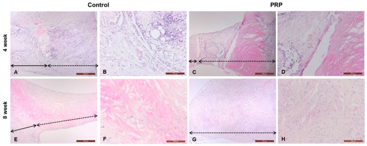
Figure 6.
Histopathological analysis of full-thickness defects of the meniscus. The defective lesions of control and PRP-treated groups were completely replaced by fibrous tissue, instead of meniscal cartilage, at four and eight weeks. PRP-treated lesions were relatively thickened with hypercellularity of fibroblasts when compared to the control (black full line arrow: repair site, black dotted line arrow: non-defective site) H & E, magnifications 40× and 100×. A 4wk (40×)-Control; B 4wk (100×)-Control; C 4wk (40×)-PRP; D 4wk (100×)-PRP; E 8wk (40×)-Control; F 8wk (100×)-Control; G 8wk (40×)-PRP; H 8wk (100×)-PRP.

