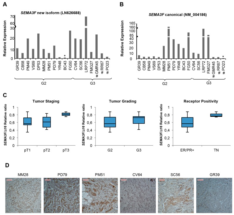
Figure 6.
Semaphorin 3F expression in BC tumor biopsies (n = 18). (A,B) qRT-PCR analysis of the new (LN626688) and the annotated (NM_004186.3) SEMA3F transcripts, respectively. Samples have been ordered according to tumor grade (G2, G3); # indicates Triple Negative biopsies; asterisks (*) indicate the sample with the lowest absolute expression, used as reference; (C) Relative ratio of ΔCt values between skipping and canonical mRNA isoforms of SEMA3F across breast cancer subtypes. TNM: pT1 (n = 7); pT2 (n = 9); pT3 (n = 2). Tumor grade: G2 (n = 11); G3 (n = 7). ER+/PR+ (n = 15); TN (triple negative samples) (n = 3); (D) Six representative images by immunohistochemistry assay on FFPE slices from breast cancer specimens are shown; N-terminal anti-Sema3F (that recognizes both the canonical and the new splicing variant) primary antibody was used. Scale Bar (in red) value = 33.76 µm.

