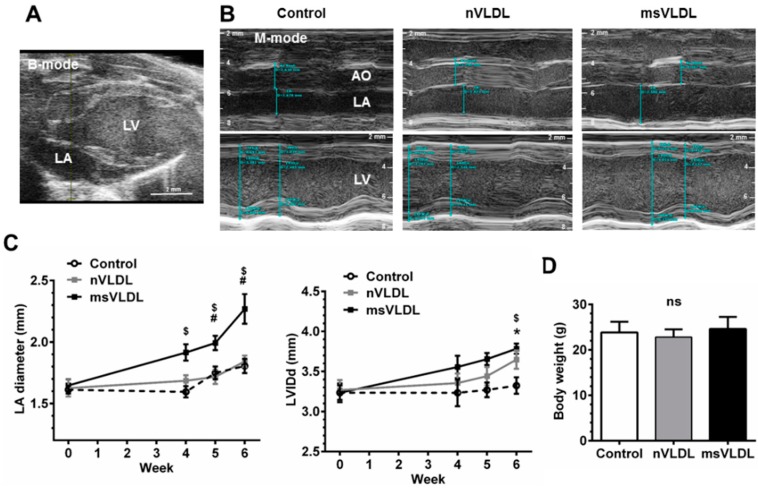
Figure 2.
Both VLDLs caused LV dilation but only MetS-VLDL caused left atrial dilation. (A) Echocardiography of murine heart. Left atrium (LA) and left ventricle (LV) were identified in B-mode; (B) M-mode images for measurements of diameters of aortic root (AO), LA and LV. LA was significantly enlarged in the MetS-VLDL injection group (msVLDL) (n = 6) but not in the normal-VLDL injection group (nVLDL) (n = 7) or the control group (n = 5); (C) Significant LA enlargement developed as early as 4–6 weeks after injection in the msVLDL group. LV dilatation developed significantly until 6 weeks. (msVLDL vs. control, $ p < 0.05; msVLDL vs. nVLDL, # p < 0.05; nVLDL vs. control, * p < 0.05); (D) No significant difference in body weight of the groups.

