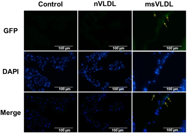
Figure 4.
Apoptosis in atrial tissue of msVLDL mice. Representative in situ terminal deoxynucleotidyl transferase (TUNEL) staining of atrial tissues from control (left), nVLDL (middle), and msVLDL (right) (n = 3 for each groups). Normal nuclei with DAPI staining appear blue. Condensed or fragmented nuclei appeared bright green and indicate cells undergoing apoptosis. Arrows indicate apoptotic atrial myocytes in the msVLDL group. The scale bars indicate 100 µm.

