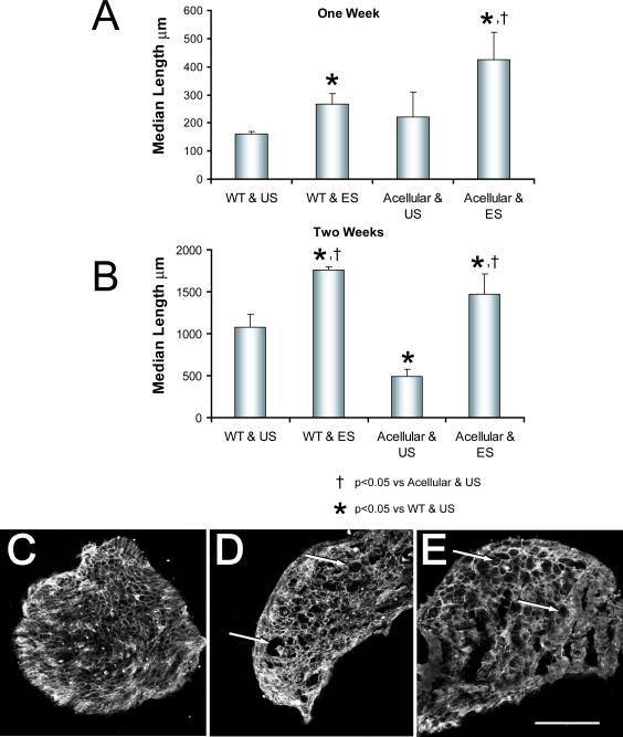
Figure 4.
Effects of electrical stimulation on the regeneration of axons through acellular grafts. The protocol is similar to that described in figure 2, except that the grafts from wild type (WT) mice used to repair the nerve either were untreated or were made acellular by repeated freeze-thawing before use. Median lengths (+SEM) of YFP+ axon profiles measured in grafts from these different donors which were used to repair electrically stimulated (ES) nerves or unstimulated (US) nerves are shown one (A) or two weeks (B) after the nerve repair. Panels C-E are images of transverse sections through acellular grafts before (C), one week (D) and two weeks (E) after being used to repair cut nerves. Each has been reacted with an antibody to laminin-2. Arrows indicate examples of enlarged spaces found in implanted grafts. Scale bar = 200 μm.

