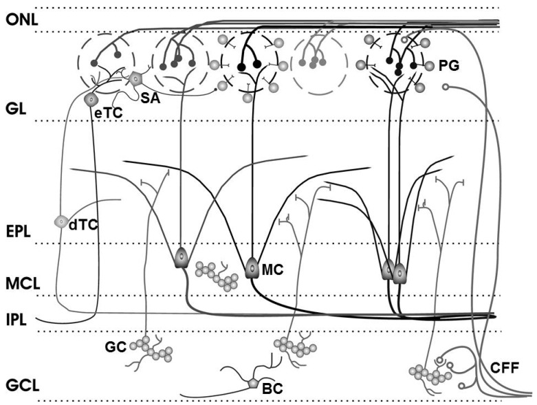
Figure 2.
Olfactory bulb circuitry. Olfactory receptor neuron axons enter the main olfactory bulb through the olfactory nerve laver (ONL), to synapse with periglomerular cells (PG), mitral cells (MC) and tufted cells (of which external, eTC, and deep, dTC, tufted cells are shown) within the glomerular layer (GL). Short Axon (SA) cell axons receive synaptic input from eTCs and form extensive interconnections between glomeruli, while mitral cell apical dendrites convey sensory information to deeper layers of the bulb. In the external plexiform layer (EPL), mitral and (deep) tufted cells extend lateral dendrites which release glutamate onto the dendrites of granule cells (GC). Mitral cell bodies are located in the mitral cell layer (MCL), which is also densely packed with granule cells. Mitral and tufted cell axons project through the internal plexiform layer (IPL) to olfactory cortex (their axon collaterals branching into the GCL are not shown). The granule cell layer (GCL) contains the major population of inhibitory granule cells. Blane’s cells (BC) within the GCL make inhibitory contact with granule cells. Centrifugal fibers (CFF) shown projecting to the GL and GCL include glutamate-releasing axons of olfactory cortex pyramidal cells receiving mitral cell output. Modified from [60]; original drawing by Cristina Shirley.

