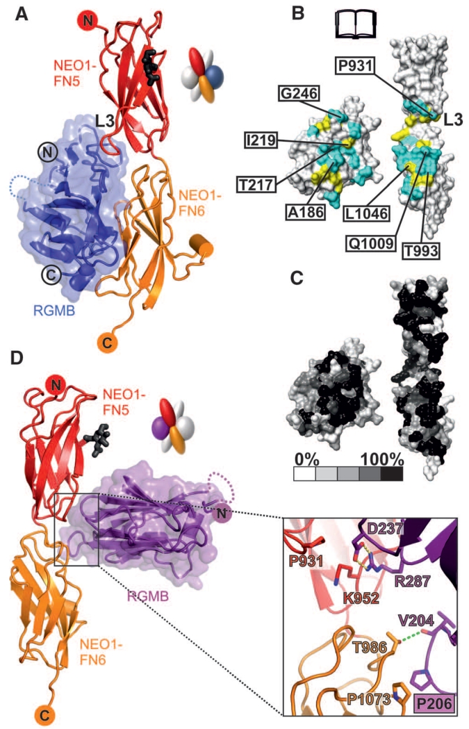
Fig. 2. Detailed interactions of the RGMB-NEO1 complex.
Color coding is as Fig. 1D. (A) Ribbon representation of the RGMB-NEO1 site-1 complex. The L3 loop of NEO1 is marked. (B and C) Open-book view showing the solvent-accessible surface of the site-1 interface (formed by 17 hydrogen bonds and 147 nonbonded contacts). (B) Interface residues (I, Ile; L, Leu; Q, Gln; T, Thr). Cyan, hydrophilic interactions; yellow, nonbonded contacts. Residues tested by site-directed mutagenesis and functional experiments are labeled. (C) Residue conservation (from nonconserved, white, to conserved, black) based on sequence alignments from vertebrate NEO1 and RGM family members. (D) Ribbon representation of the RGMB-NEO1 site-2 complex. The site-2 interaction uses the RGMB β5-β6 and β10-β11 loop regions contacting the NEO1 FN5 and FN6 domains. K, Lys; V, Val.

