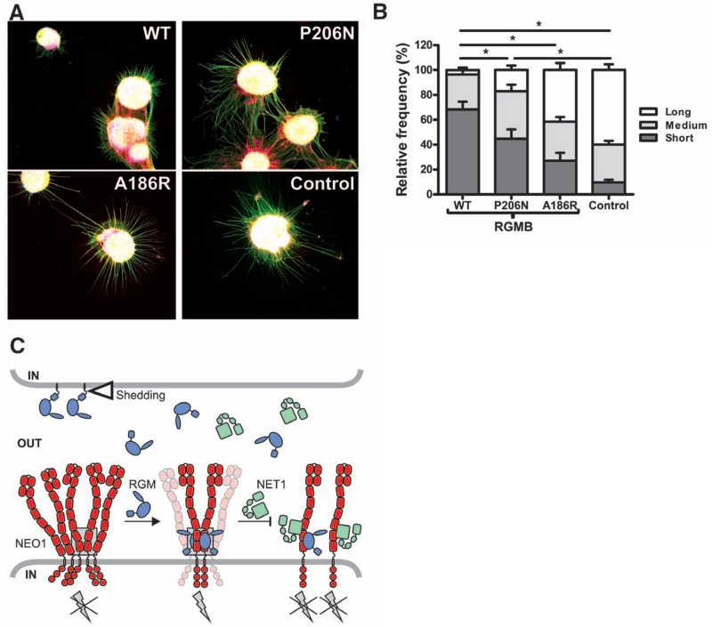
Fig. 4. Functional analysis of site-1 and -2 mutations on RGMB neurite growth inhibitory effects.
(A) Representative examples of P9 mouse CGN explants on coverslips coated with RGMB-WT (top left), site-2 mutant RGMB-P206N (top right), site-1 mutant RGMB-A186R (bottom left), or control (bottom right) proteins. Green, βIII-tubulin; red, F-actin; blue, nuclei. (B) Quantification of CGN neurite outgrowth. Distribution of neurite length (short, medium, and long) relative to the control is displayed (total number of explants analyzed for WT, n = 27; P206N, n = 24; A186R, n = 26; control n = 23); error bars are SEM, and *P < 0.01. (C) Model of trans RGMB-NEO1 signaling. RGM ectodomains can be shed by proteolytic or phospholipase activity (open triangle). RGM-binding to preclustered NEO1 results in formation of NEO1 dimers with a defined, signaling-compatible orientation that may be part of a supramolecular clustered state. This arrangement leads to activation of downstream signaling via RhoA (9) (gray lightning bolt). NET1 can inhibit RGM signaling by either simultaneous NEO1 binding or competing with the RGM-NEO1 interaction. The gray box highlights the RGM-NEO1 signaling hub observed in the crystal structure.

