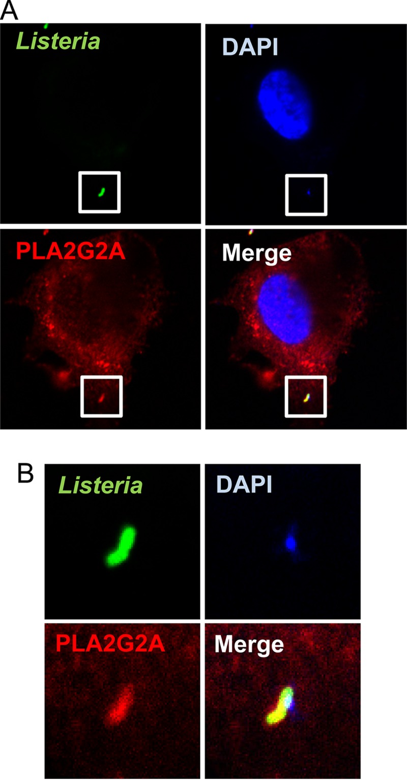
FIG 5.

Intracellular colocalization of PLA2G2A and L. monocytogenes in HepG2 cells. IL-22-treated HepG2 cells were washed to remove PLA2G2A in supernatants, infected with L. monocytogenes, and incubated for 1 h. Extracellular bacteria were washed out, fixed, permeabilized, and stained to detect intracellular PLA2G2A and L. monocytogenes. The cells were mounted with medium containing DAPI to visualize cellular and bacterial DNA. The cells were observed by confocal microscopy with a 60× objective and 4× zoom (A). The insets (A) are magnified in panel B to show colocalization of PLA2G2A, L. monocytogenes, and DNA. The images show a cell with intracellular bacteria that is representative of three independent experiments.
