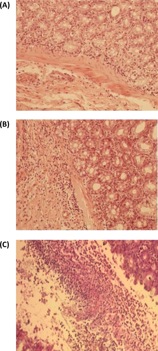
FIG 5.
Cecum histology of hamsters surviving spore challenge after prophylactic treatment with L. paracasei BL23 strains displaying cell wall-anchored VHH fragments neutralizing TcdB. Hematoxylin- and eosin-stained sections of ceca from different treatment groups were assessed for inflammation and cellular destruction. (A) Normal cecum mucosa of hamster 54-5, with no signs of lesions or inflammation. (B) Mild colitis, with lymphocyte and histiocyte infiltration (grade 2), in the mucosa of the cecum of hamster 54-2. (C) Severe colitis, with necrotic masses with fibrin, macrophages, and neutrophils (pseudomembranes) (grade 5), in the mucosa of the cecum of a nonprotected hamster challenged with C. difficile TcdA− TcdB+ spores.

