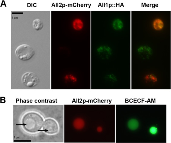
FIG 4.

(A) All2p-mCherry fusion proteins localize in the cytoplasm, with accumulation in vacuoles. For coexpression studies, single cells expressing All2p-mCherry (red) and All1p::HA (green) fusion proteins were grown in YNB for 16 h at 37°C and observed by using differential interference contrast microscopy and fluorescence. The imaging revealed the cytoplasmic location and colocalization of both fusion proteins. (B) All1-mCherry fusion proteins were also found to colocalize in yeast vacuoles by phase-contrast microscopy (indicated by an arrow) and BCECF-AM staining (scale bar, 5 μm).
