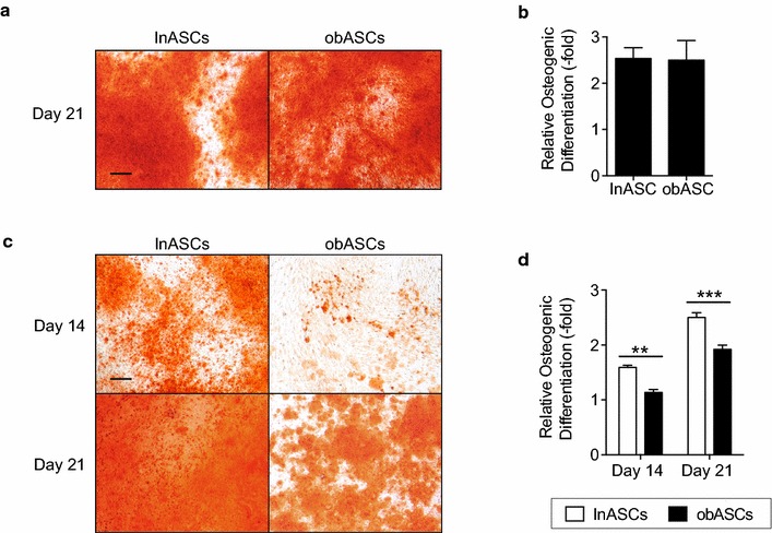
Fig. 1.

obASCs display reduced osteogenic differentiation in an estrogen depleted environment. a lnASCs (N = 6 donors) and obASCs (N = 6 donors) were cultured in ODM and stained with alizarin red after 21 days for calcium deposition. b To quantify the amount of alizarin red staining, stains were eluted with CPC and optical density was read. c lnASCs (N = 6 donors) and obASCs (N = 6 donors) were cultured in CDS-ODM and stained with alizarin red after 14 and 21 days for calcium deposition. d To quantify the amount of alizarin red staining, stains were eluted with CPC and read at 590 nm. Scale bar represents 200 µm. Bars, ± SEM. **, P < 0.01; ***, P < 0.001
