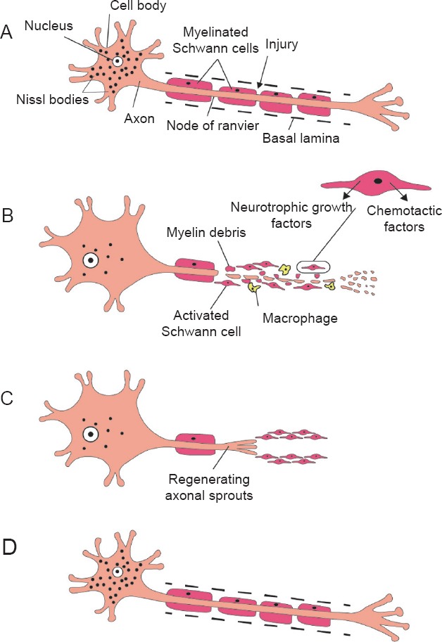
Figure 1.

Wallerian degeneration in peripheral nervous system and axonal regeneration.
A typical motor neuron with its axon surrounded by myelin sheath formed by Schwann cells (A). After injury, the distal detached nerve segment degenerates, and the surrounding myelin breaks down. The proximal segment of the axon undergoes degeneration that extends proximally only as far as the first node of Ranvier. The proximal segment undergoes chromatolysis; the Nissl bodies dissolve, the nucleus migrates towards the periphery of the cell and the size of the neuronal cell body and nucleus increase. Schwann cells become activated; they rapidly demyelinate, proliferate and dedifferentiate to a more progenitor-like cell type. Schwann cells also induce chemotactic migration of macrophages and secrete several neurotrophic factors needed for regeneration. Axonal and myelin debris are phagosytosed by both Schwann cells and recruited macrophages (B). The proliferating Schwann cells form a line of cells called bands of Büngner within their basal lamina. These bands guide axonal sprouts during regeneration (C). In the later stages of regeneration, Schwann cells redifferentiate into myelinating cells and evidence of chromatolysis is no longer present (D).
