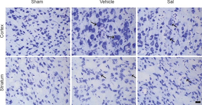
Figure 2.

Salidroside improved the morphology of neurons in the cortex and striatum after ischemia /reperfusion (Nissl staining, × 400).
Histological changes in the cortex and the striatum were evaluated by Nissl staining. In the vehicle group (vehicle + MCAO), cells in the cortex and striatum were disarranged and a large number of cells were swollen. Cellular shapes changed from triangular to rounded, and normal cell structures were invisible. In the Sal group (salidroside + MCAO), the morphology of neurons was regular. Arrows show swelling and distension. Sham: Sham operation group; vehicle: 0.9% saline-treated group; Sal: 30 mg/kg salidroside group; MCAO: middle cerebral artery occlusion. Scale bar: 20 μm.
