Figure 4.
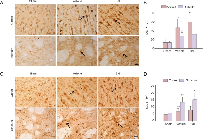
Effects of salidroside on Nrf2 and HO-1 immunoreactivity in the cortex and striatum of rats with cerebral ischemia (× 400).
Immunohistochemical staining was used to detect the distribution of Nrf2 and HO-1 in cortex and striatum at 24 hours after cerebral ischemia and reperfusion. (A) Representative images of immunohistochemistry for Nrf2. Brown Nrf2-stained cells were prominently observed in the vehicle group. In the cortex, the Nrf2 immunoreactivity was enhanced in the Sal group compared with the vehicle group. Arrows show Nrf2-immunoreactive cells. (B) Quantification of the IOD for Nrf2. (C) Representative images of immunohistochemistry for HO-1. Brown HO-1-stained cells were prominently observed in the vehicle group. In the striatum, the HO-1 immunostaining intensity was enhanced in the Sal group compared with the vehicle group. Arrows show HO-1-immunoreactive cells. (D) Quantification of IOD for HO-1. Data are expressed as the mean ± SD (n = 5; one-way analysis of variance, followed by the least significant difference post hoc test). **P < 0.01, vs. sham; #P < 0.05, vs. vehicle. Sham: Sham operation group; vehicle: 0.9% saline-treated group; Sal: 30-mg/kg salidroside group; IOD: integrated optical density; Nrf2: nuclear factor erythroid 2-related factor 2; HO-1: heme oxygenase-1. Scale bars: 20 μm. Experiments were conducted in triplicate.

