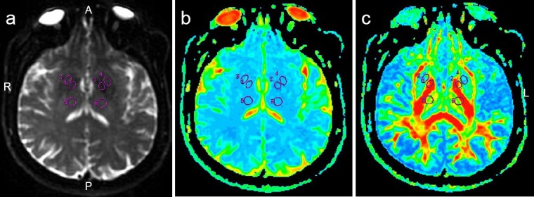
Figure 1.

Regions of interest in a female Parkinson's disease patient (63 years old; Hoehn and Yahr Scale stage 1.5).
(a) T2-weighted image; (b) ADC map; (c) FA map. 1–6 refer to regions of interest on FA and ADC maps. A: Anterior; P: posterior; R: right side; L: left side; ADC: apparent diffusion coefficient; FA: fractional anisotropy.
