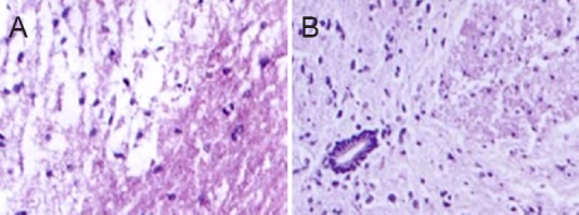
Figure 2.

Histomorphological changes in the injured spinal cord 2 weeks after electroacupuncture (hematoxylin-eosin staining, × 200).
(A) Control group: spinal cord edema with multifocal hemorrhage, indicating successful model establishment. (B) Electroacupuncture group: many neurons survived with only light swelling and limited necrosis, with a tight arrangement. Karyocytes with a small size were seen.
