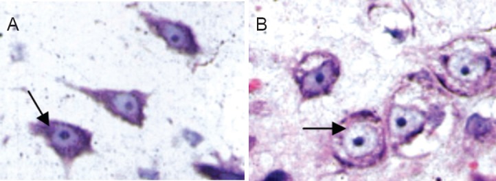
Figure 4.

Effects of electroacupuncture on the morphology of Nissl bodies in motor neurons in rats with spinal cord injury (thionine-giemsa staining, light microscope, × 1,000).
At 4 weeks after injury, Nissl bodies were massive and abundant in the electroacupuncture group (arrow, A). Few Nissl bodies, lightly stained, were visible in the control group (arrow, B).
