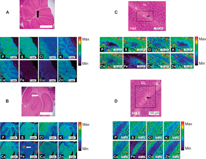
Fig. 3. XFM analysis of the elemental alterations in the cerebellum during murine CM.

(A and B) XFM elemental maps (P, S, Cl, K, Ca, Fe, Cu, and Zn) collected with a 5-μm step size of the cerebellum of control (A) and CM (B) mice. H&E histology is presented for the entire cerebellum and at sites of healthy and hemorrhaged vasculature. IL, inner layer (white matter); GL, granular layer (gray matter). Arrows indicate the location of healthy vasculature in controls (black) and hemorrhaged (white) vasculature in ECM. (C and D) High–spatial resolution XFM elemental maps (P, S, Cl, K, Ca, Fe, Cu, and Zn) collected with a 0.5-μm step size of the cerebellum of control (C) and CM (D) mice. H&E histology is presented for the sites of healthy and hemorrhaged vasculature, indicated by black arrows. Note that high relative Fe content is only observed within the microvessel in the sham animal (similar to the preliminary investigation, fig. S8), whereas high Fe is observed throughout the white matter in the CM mouse, with low Fe observed in the white matter surrounding the vessel. IL, inner layer (white matter); GL, granular layer (gray matter).
