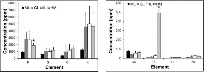
Fig. 4. XFM elemental concentrations showing a significant difference in the P and Fe concentrations observed in hemorrhaged white matter tissue in CM mice, relative to healthy molecular layer, granular layer, and white matter tissue in control mice (n = 5).

*Significant difference relative to control tissue. IL, inner layer (white matter); GL, granular layer (gray matter); ML, molecular layer; HM, hemorrhaged tissue.
