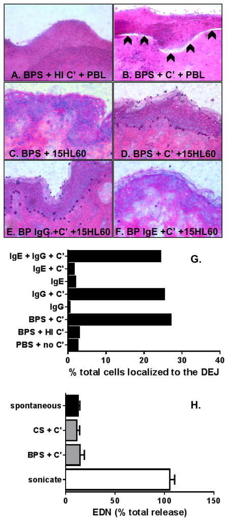
Figure 4. IgG and complement are required for eosinophil localization to the BMZ.
Cyrosections of human skin were incubated with BP sera (BPS) + heat inactivated (HI) or fresh complement (C′) + PBL from a healthy donor, as negative (A) and positive controls (B), respectively. Slides were stained with H & E, images were captured via light microscopy. A subepidermal split is indicated by the arrowheads (B). Next, cryosections were incubated with BPS + 15HL60 eosinophils ± complement (C, D). Incubation of cryosections with purified IgG or IgE + C′ + 15HL60 cells revealed that eosinophil localization to the BMZ is dependent on IgG and C′. The %of the total overlaid cells that localized to the DEJ was determined using NIH Image J (G). Collection of supernatants from cryosection experiments revealed that 15HL60 eosinophils do not degranulate under these conditions (H). Likewise, application of 15HL60 lysates did not cause a subepidermal split (data not shown). Bars represent the average value from two cryosections treated identically (G) or as mean ± SD of supernatants collected from triplicate samples treated identically (H).

