
Figure 5.
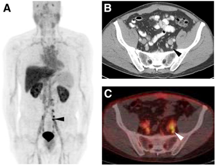
18F-DCFBC PET maximum-intensity projection (A), axial CECT (B), and axial fused 18F-DCFBC PET/CT (C) images demonstrating intense 18F-DCFBC uptake in multiple small pelvic lymph nodes that had been deemed too small to be definitively disease-involved on CECT (black and white arrowheads in A–C). Lymph nodes decreased in size on follow-up imaging and correlated with fall in patient's prostate-specific antigen level to undetectable.