
Figure 6.
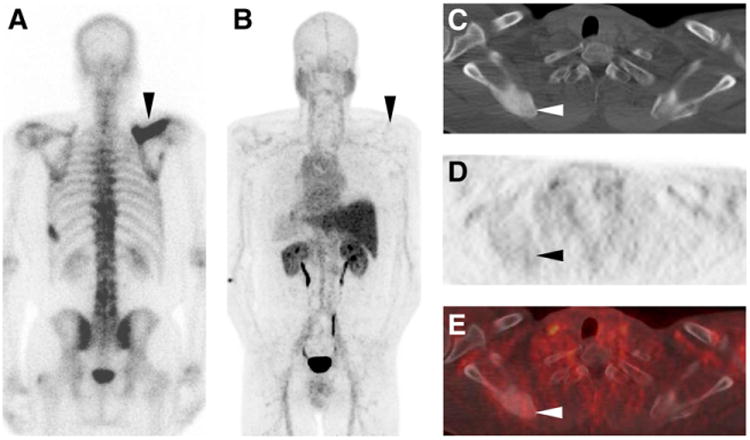
Posterior projection planar BS (A), 18F-DCFBC PET maximum-intensity projection (posterior view, B), axial CT (C), axial 18F-DCFBC PET (D), and axial fused 18F-DCFBC PET/CT (E) images from patient who was postprostatectomy with rising prostate-specific antigen and was naïve to systemic androgen-deprivation therapy and chemotherapy. Imaging demonstrates intense 99mTc-MDP uptake on BS and corresponding dense sclerosis on CT of right scapula without significant 18F-DCFBC uptake (black and white arrowheads in A–E). This lesion progressed in extent to involve more of scapula on follow-up imaging in correlation with rising prostate-specific antigen level in this patient.