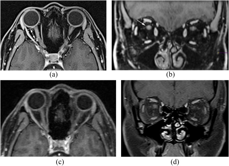
Figure 6.
Post-contrast (a) axial and (b) coronal radial-volume-interpolated breath-hold examination (Radial-VIBE) images demonstrate relatively sharply defined segmental enhancement (double headed arrow) of the intraorbital portion of the right optic nerve (arrow) consistent with suspected optic neuritis. In comparison, (c) post-contrast axial three-dimensional magnetization-prepared gradient echo with water excitation and (d) post-contrast coronal two-dimensional T1 turbo spin echo with fat suppression images demonstrate subtle asymmetric enhancement of the right optic nerve.

