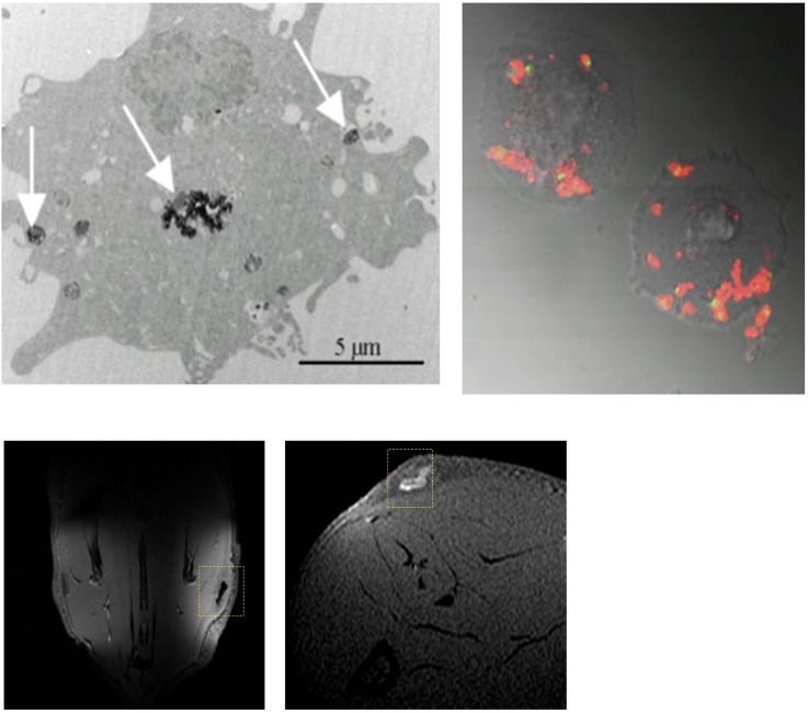
Figure 1.
Nanoparticle labelling and imaging of cells. Top panels: an electron microscopy (left) and fluorescent microscopy (right) image of human umbilical vein cells labelled with iron oxide nanoparticles and fluorescent Gd–liposomes, respectively, showing intracellular presence of the nanoparticles after labelling procedure. Arrows indicate intracellular deposits of iron oxide nanoparticles. Bottom panels: magnetic resonance images obtained from rats injected subcutaneously with cells labelled with iron oxide particles or Gd–liposomes (liposomes containing gadopentetate dimeglumine in the water phase).

