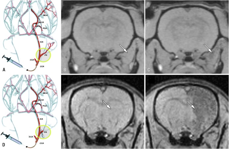
Figure 6.
Real-time monitoring of injection accuracy with MRI. (a) Diagram of procedure with pterygopalatine artery left intact. After ligation of external carotid and occipital arteries, common carotid artery was cannulated and SPIO-labelled cells were infused. (b, c) T2* weighted MR images of rat brain and surrounding muscles obtained immediately before (b) and after (c) injection demonstrate that vast majority of cells are localized into extracerebral tissue (arrows), with negligible binding within brain. (d) Diagram of procedure with ligation of pterygopalatine artery. All infused cells were perfused into internal carotid artery and localized successfully into ipsilateral hemisphere. (e, f) MR images obtained immediately before (e) and after (f) injection. Arrows indicate area of cell docking. CA, choroidal anterior artery; CCA, common carotid artery; ECA, external carotid artery; ICA, internal carotid artery; MCA, middle cerebral artery; PA, pterygopalatine artery; PCA, posterior cerebral artery. Reprinted from Gorelik et al132 with permission from the Radiological Society of North America.

