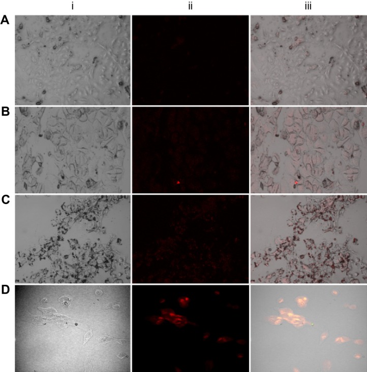
Figure 9.
Confocal microscopic images of Mn:ZnS-treated MCF-7 cells (A), FACS-Mn:ZnS-treated MCF-7 cells (B), MDA-MB-231 cells (C), and MCF-10 cells (D), respectively.
Note: From each zone, (i) represents the normal transmission image, (ii) represents the fluorescence image, and (iii) represents the combination/overlaying of both transmission and fluorescence images of the corresponding cells.
Abbreviations: FACS-Mn:ZnS, folic acid–chitosan stabilized Mn2+-doped ZnS; ZnS, zinc sulfate.

