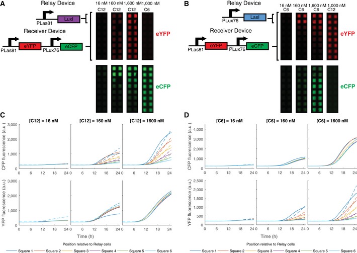
Figure 4. Relay devices respond to one HSL signal by sending the other.

-
A, BImages at t = 1,500 min of chromosomal constitutive mRFP1 cells transformed with the pR33S175 double receiver and a relay device plated in 2 rows of 2 grid squares adjacent to cells containing the pR33S175 double receiver plus empty vector (for antibiotic resistance) in wells containing AHLs at the concentrations specified. (A)The pLas81LuxI relay device. (B) The pLux76LasI relay device.
-
C, DeCFP (top) and eYFP (bottom) fluorescence (arbitrary units) plotted against time for the grid squares containing receiver plus empty vector. Solid lines plot data while dashed lines plot the model. Position relative to the grid squares containing relay devices are according to the color code shown. (C) The pLas81LuxI relay device. (D) The pLux76LasI relay device.
Source data are available online for this figure.
