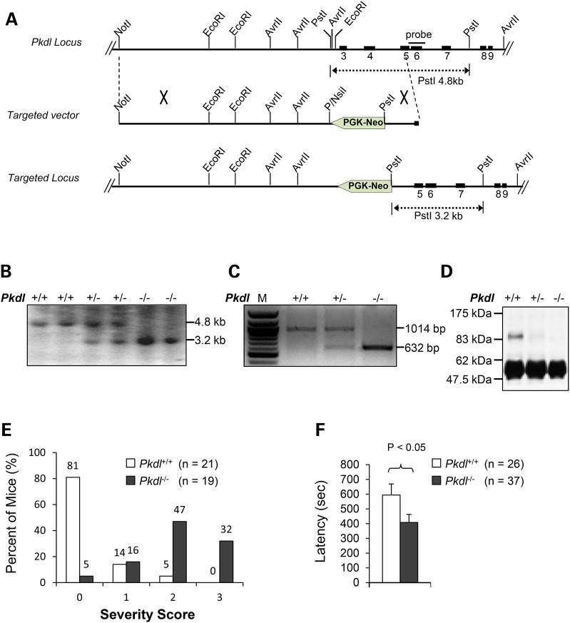
Figure 2.
Pkdl−/− mice are sensitive to PTZ. (A) Targeting strategy for Pkdl and restriction map of the Pkdl gene. The solid boxes represent the exons of the Pkdl gene and the interconnecting lines indicate introns. The probe used for Southern blotting is shown as a bar. (B) Southern blot analysis of mouse tail DNA. The 3.2 and 4.8 kb bands represent the germ-line and targeted allele, respectively. (C) RT-PCR analysis of total RNA extracted from mouse brains using primers mPcLf605 5′ACACAGCCGAGAACAGGGAGCTT3′ and mPcLr611 5′ GCATACGTGTCTGGCTGTTGCAG3′. No normal Pkdl mRNA was detected in the brains of adult Pkdl−/− mice by RT-PCR analysis, but a truncated Pkdl mRNA was present. M, 100 bp DNA marker. (D) Immunoblotting analysis of the PCL protein. Anti-PCL antibody is specific for the N-terminal portion of the PCL protein. Testis extracts from Pkdl+/+, Pkdl+/− or Pkdl−/− mice were immunoprecipitated with anti-PCL and immunoblotted with the same antibody. PCL protein was not detected in Pkdl−/− mice. (E) The severity of seizure increases in KO mice when compared with WT mice (PTZ = 40 μg/g). 0: no response; 1: isolated twitches; 2: tonic-clonic convulsions; 3: tonic extensions and/or death. (F) Latency in KO mice is reduced compared with WT littermates (PTZ = 50 μg/g).

