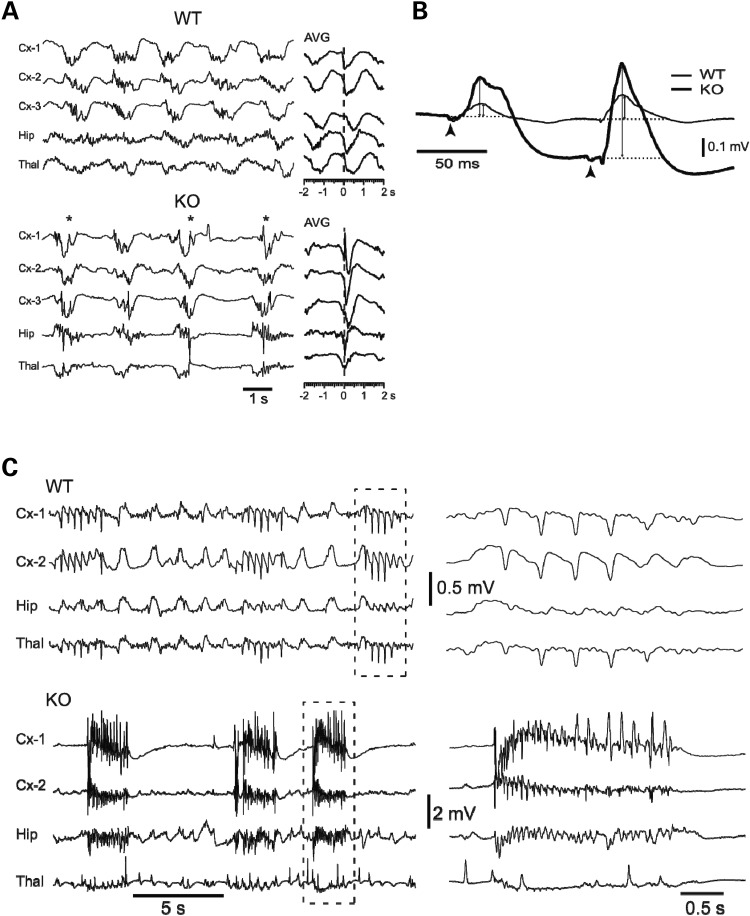
Figure 3.
LFP recordings suggest a loss of control of excitability in Pkdl−/− mice. (A) Under ketamine–xylazine anesthesia, EEG recording of spontaneous activity from neocortex (Cx-1, -2 and -3), hippocampus (Hip) and thalamus (Thal) showed the slow oscillation (WT = 0.6 Hz; KO = 0.4 Hz) synchronized among the three structures as shown in the average (AVG, n = 20 cycles) centered on negative peaks of Cx-1. KO showed high-amplitude spikes. (B) Responses to thalamic stimulation of ventrobasal (VB) nucleus of the thalamus. (C) Seizure threshold is lower in KO mice. PTZ (40 μg/g) triggered mild spike–wave seizures in WT mice (n = 6), while KO mice (n = 10) showed severe tonic-clonic seizures.

